When people talk about forensic, I always think of NCIS (because that is one of my favourite tv shows on Earth). And mehh okay maybe CSI as well but I don't really like CSI as much. Never mind that.
So we gathered inside the seminar room where they do their presentations and the head of forensic pathologist (crap I don't remember her name) came and introduced herself. What I can remember was that there are only 3 pathologists to handle all forensic cases in Johor, which is a really sad thing. Pathologists are highly sought after you know. My lecturer once joked that there are only 2 people who know about what the patient has - one is God and the other is the pathologist. Even when you become a hotshot medical officer working in a ward, you may make a diagnosis based on what you see or examine on the patient but you would still need a pathologist to confirm your diagnosis. Pretty humbling, huh?
Okay then the doctor told us that we are lucky today because there is a new case, so she wanted us to participate in this today's autopsy. She wanted to know our differential diagnoses of the case and so she presented us the case (as told by her husband):
- 36 year-old Malay woman
- Nulliparous (never given birth), married for 14 years
- Past history of hypertension
- Been complaining of headache, shortness of breath and chest pain at nights
- For the last week she was feeling lethargy as well
- During a wedding reception she felt unwell and went to clinic where she died there at 6 pm yesterday (from police report)
- Her body was brought to the hospital around 9.30 pm last night and schedule for autopsy today at 10 am.
Because her symptoms include chest pain, shortness of breath and tiredness, I made my differentials as follow:
- Heart failure
- Pulmonary embolism
- Ischaemic heart disease (IHD)
Suffice to say, all my differentials are cardiovacular in origin, because if the patient has chest pain, I strongly believe that cardiovascular is the problem unless proven otherwise.
Okay, after that we were brought to the autopsy. Jeng jeng jengggg...
And there she was.
On the metal autopsy table, covered with white cloth.
When she was revealed, she was still in her clothes (blue jeans, purple shirt). Her hands were tied together for reason I believe to prevent them from falling or hanging over when they transferred her here.
And that is the first time I saw a dead person.
The reality is always there around me. I mean I know that people live and people die. Even in the hospital we generally see alive people. Sick, yes, deathly ill, sometimes, but alive and still kicking. To see one in front of you, to me, put me into place.
At first it was like a raw feeling. That yes, that is a dead person. Yes, sometime in the future you will be dead as well. Will I become like her, then? Or much better? Or worse?
But this morning, all of these thoughts that you are reading now were not expressed. I stood there, calmly listened to the doctor's explanation about what happened to the body when they died.
Okay, some explanations:
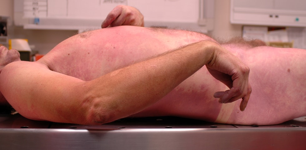
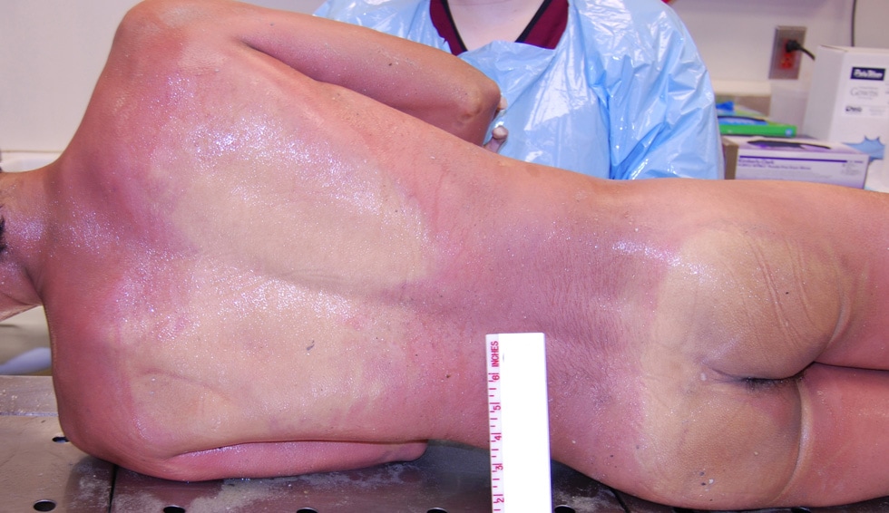
- blood collects in the most dependent parts of the body (livor mortis)
- the body stiffens (rigor mortis)
- the body begins to cool (algor mortis)
And she was VERY VERY cold to touch. Like metal-steel-in-a-fridge kind of cold. It was a surprise to me but considering all this experience is new to me everything then is a surprise. She has a patch of baldness on her head, she was obese (estimated weight of 100kg), there were no bruises, there was froth on her nostrils, and there was some blood in her mouth but I can't be sure about that.
Then the gruesome part of autopsy began:
First is the Y-shaped incision on the chest. Here is the kindest picture available on the Internet:

After that they cut the rib cages and sternum because they wanted to take out the heart. But before they took out the heart, they had to take a blood sample first. Blood sample can be taken from a corpse within the first 8 hours after death. After that the blood will be contaminated by the bacteria living in the gut.
The way they took the blood sample is by searing. They heated the scalpel with fire (ideally using a Bunsen burner) in order to sterile the scalpel. Then they made a cut onto the right atrium (blood away from the body) and immediately after they used a syringe to draw out the blood. Then they will send the blood to the lab for analysis.
One of the doctors there named Dr Yazid and he taught us heart anatomy there and then. He showed where are the vena cavas and the arteries etc. Now I know that the normal weight of a human heart is:
- for man, 0.45% of total body weight
- for women, 0.4% of total body weight
If the human heart weighs more than percentage of their body weight, we can say that they have an enlarged heart (cardiomegaly).
The reason in autopsy they take out all organs is to determine the cause of death. They took out the heart because they suspect the heart is the problem. Dr Yazid traced out the coronary arteries and then made small vertical cuts along the horizontal heart muscles where the arteries lie within. All cuts are approximately 0.5cm apart.
The reason he did that because he wanted to know where along the artery the blockage is. Coronary heart disease is a condition when the coronary arteries (the blood vessels that supply the heart) are blocked or occluded. Dr Yazid found that a small length (less than 5mm long) of the left artery descending is occluded (up to 70% occlusion).
At the same time Dr Yazid was teaching us about the heart, the other doctors were busy cutting her skull open (but before that, they pulled down the scalp over her face. Try to imagine that because any pictures put here will make you off your appetite for the rest of the week hahaha).
Then they removed the brain and again we learned about the anatomy of the brain. From the autopsy, the brain was intact with no signs of bleeding (haemorrhage) or tissue death (ischaemic or necrosis).
Of these two organs, there is a few things that struck me:
- Coronary arteries are wayyy smaller than I realised. No wonder a lot of people clogged up their arteries and died from a heart attack.
- Brain is much smaller than what I had in mind. (Oh God this is not a pun I swear I don't mean it)
After heart and brain removal, they washed the organs and weighed them. Then they sliced them into many segments. Yep, like the butchers you see in the market or during Raya Korban. Straight out hardcore stuff. They wanted to know if there is something wrong in the organs so yeahh they strongly reminded me of Hannibal Lecter.
Okay, nearing the end now (of the post, not of the autopsy)
The idea of autopsy is to determine the cause of death. To do so they have to remove all vital organs. After heart and brain, they removed all abdominal organs. That include lungs, liver, pancreas, spleen, kidneys, guts, ovaries, and uterus. You guessed correctly that at this point the smell was very hard to ignore because the gut is where all the yucky part resides. Besides that, in autopsies they will also take out tissue samples and see under the microscope for any histology changes. It means that they can figure out problems at microscopic level. You also already know that fluids are also sampled and sent to the lab for all kinds of analysis.
Long story short, (well I'm kinda feel tired of typing this long anyway hahaha) here are the findings:
- occluded coronary artery
- cardiomegaly
- hepatization of lungs (the lungs become firm like the liver; they are supposed to feel like sponge)
- hepatomegaly (enlarged liver, 3.2kg)
- spleenomegaly (enlarged spleen)
- haemorrhagic pancreatitis (the pancreas is a bloody mess, like literally a bloody mess)
- renal cortical necrosis (the kidneys are scarred, probably due to her hypertension)
- multiple ovarian cysts (together with her facial hair - hirsutism - indicative of PCOS)
- excoriation of uterus
After that because we were pressed for time we have to leave the doctors to do their autopsy and we had to catch the van back to campus.
So, my take on today visit to forensic autopsy?
Bloody scary, bloody messy.
Gruesome, scary, and yep bloody.
Understaffed, under appreciated, undervalued,
Informative, important,
It's an evidence medicine to determine the cause of death.
I should explain more about the two forms of autopsy; the medicolegal and the clinical ones.
But to summarize, medicolegal involved the police (homicde, suicide, unnatural death, etc.) and clinical one is.. without the police.
I am so not going to be a pathologist or a forensic one for that matter.
God help me.


This comment has been removed by the author.
ReplyDelete